New classification system for brain tumours
Doctors at Universitätsklinikum Erlangen have developed a simple radiological method to predict the development of gliomas
Despite modern chemoradiation therapy it is still very difficult to give reliable prognoses for malignant gliomas. Surgical removal of the glioma is still the preferred method of treatment. Doctors at Universitätsklinikum Erlangen’s Department of Neurosurgery have now developed a new procedure for analysing radiological imaging scans which makes it possible to predict the course of a disease relatively precisely. Their findings have now been published in the journal ‘Scientific Reports’.*
A quick and safe way of classifying a tumour’s operability
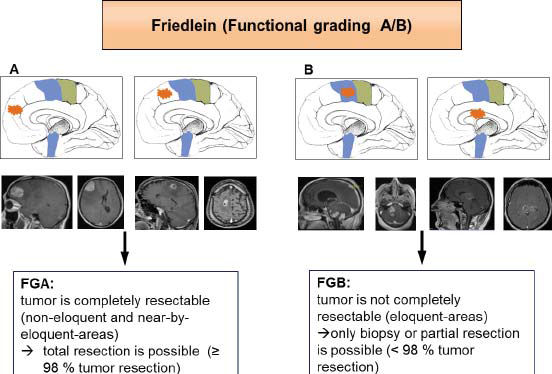
The Friedlein Grading A/B (FGA/B) classification system – named after the physician Katharina Friedlein – is a quick and precise way of determining whether surgical removal is the best possible treatment method for a given tumour. Essentially, the Erlangen-based doctors classify tumours according to their position in the brain in the context of a routine magnetic resonance imaging (MRI) scan. Tumours that are not located in functional brain regions or that are located at a certain distance from such regions are classified as FGA, while tumours that are close to or inside a functional brain region are classified as FGB.
Developing precise low-risk strategies
With the FGA/B method it possible to plan the consequences of tumour surgery, which is crucial for the success of the treatment, in a precise, low-risk and quantitative manner. This makes the Friedlein Grading system the first classification system which can be easily applied in clinical practice. ‘There have already been several attempts in medicine to develop such a classification system. However, most approaches were too complicated and were based on academic values only, which made it difficult to use them in clinical practice,’ says PD Dr. Nicolai Savaskan from FAU’s Chair of Neurosurgery. ‘The FGA/B method can be applied on the basis of a standard MRI scan which glioma patients have to undergo anyway and is highly reliable despite being so simple. We hope that our colleagues in neurosurgery departments in smaller hospitals will also be able to use it successfully in everyday clinical practice.’
Contact the cancer information service if you have any questions
The researchers evaluated their new method in a clinical trial which has recently been published. For more information, contact the cancer information service at the Comprehensive Cancer Center Erlangen-EMN (CCC) via the free hotline +49 800 8510085 or krebsinformation@uk-erlangen.de.
*Scientific Reports, July 2015. A new functional classification system (FGA/B) with prognostic value for glioma patients. Katharina Friedlein, Yavor Bozhkov, Nirjhar Hore, Andreas Merkel, Björn Sommer, Sebastian Brandner, Michael Buchfelder, Nicolai E. Savaskan & Ilker Y. Eyüpoglu
Further information:
PD Dr. Nicolai Savaskan
Phone: +49 9131 8544748
nicolai.savaskan@uk-erlangen.de