
FAU researchers take a leading role in international study – new findings could contribute to new approaches for therapy
NMOSD is an inflammatory autoimmune disorder that damages support and supply cells in the central nervous system, known as astrocytes. Researchers from Friedrich-Alexander-Universität Erlangen-Nürnberg (FAU), Uniklinikum Erlangen, LMU University Hospital Munich, the Medical University of Vienna and other international research institutions have now identified an auto-antibody that might be involved in triggering NMOSD-like diseases. It attacks the astrocyte protein MLC1 that regulates fluid and electrolyte homeostasis in the brain. The study, that has been published in the journal “Science Translational Medicine” increases our understanding of autoimmune diseases affecting the central nervous systems and lays the foundations for new treatment concepts.
Neuromyelitis optica spectrum disorder (NMOSD) is a rare autoimmune disease of the central nervous system in which the immune system attacks structures in the brain, the spinal cord and the optical nerve. It mostly affects astrocytes, glial cells that have important support and supply functions in the nerve tissue. As early as 2005, the majority of NMOSD patients were shown to have an antibody that attacks the astrocyte protein aquaporin-4 in their serum. “This fundamental discovery indicated that astrocytes are a key target of the autoimmune attack,” says Prof. Dr. Simone Mader, head of Translational Immunology at Uniklinikum Erlangen. Initially, the discovery helped distinguish NMOSD from multiple sclerosis (MS) and other related diseases with similar symptoms.
However, AQP4 antibodies cannot be found in all patients with NMOSD or symptoms similar to NMOSD. This raises the question of whether further antibodies are involved in triggering these diseases. “Particularly in these seronegative cases, it has remained largely unclear which molecular mechanisms are behind the disease,” explains Mader.

“The findings suggest that the studied auto-antibodies are not merely an accompanying phenomenon but are involved in the processes that cause the disease.”Prof. Dr. Simone Mader
MLC1 auto-antibodies may trigger NMOSD-like disease
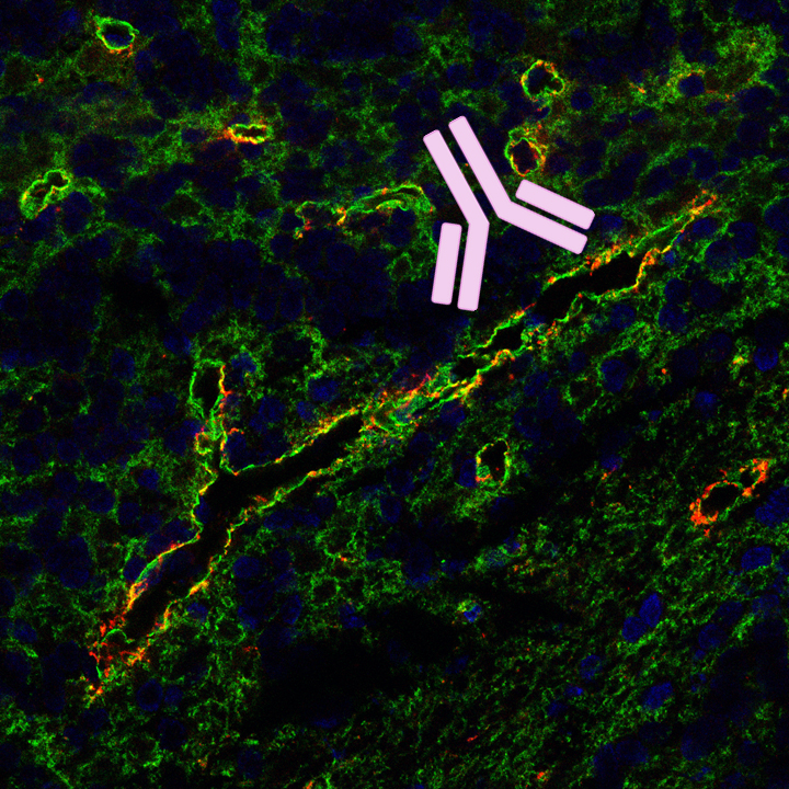
In an international research collaboration, a team led by Simone Mader identified an auto-antibody that also attacks astrocytes and could trigger NMOSD-like diseases. “The starting point was a characteristic antibody staining pattern that we observed when examining brain tissue sections with the serum of a patient,” says Mader. Through a targeted search for the underlying target protein, MLC1 was identified as the autoantigen: a membrane protein that regulates fluid and electrolyte homeostasis as well as cell volume in the brain and spinal cord.
Building on this, the researchers developed a cell-based test to detect auto-antibodies against MLC1. The test is based on a principle that has already proven effective in detecting auto-antibodies against other membrane proteins such as aquaporin-4. The result: Among patients with inflammatory diseases of the central nervous system, the researchers identified four individuals with MLC1 auto-antibodies; none of whom tested positive for aquaporin-4 antibodies. Further laboratory investigations showed that MLC1 antibodies bind to astrocytes, damaging or even destroying them completely. Simone Mader: “The findings suggest that the studied auto-antibodies are not merely an accompanying phenomenon but are involved in the processes that cause the disease.“
Goal: To improve the classification of neurological autoimmune diseases
The joint study also involved the research groups of Prof. Dr. Edgar Meinl and Prof. Dr. Naoto Kawakami, as well as the neuroimmunology outpatient clinic headed by Prof. Dr. Tania Kümpfel at LMU University Hospital Munich, the research group of Prof. Dr. Monika Bradl at the Medical University of Vienna, and numerous other research partners. The findings expand the understanding of NMOSD and NMOSD-like autoimmune diseases. They show that even in patients where the known AQP4 antibodies are not detectable, an antibody-mediated immune response against astrocytes may be present.
In further studies, MLC1 antibodies will now be examined in larger patient groups. “Identifying additional disease-relevant auto-antibodies could help more precisely distinguish between the different forms of neurological autoimmune diseases,” says Simone Mader. “In the long term, such insights could also provide starting points for more targeted therapies.”
“Antibodies against MLC1 found in patients with NMOSD-like disease mediate astrocytopathy in rodent models”
*DOI: 10.1126/scitranslmed.ady0403
The current study exemplifies a key research focus of Translational Immunology at Uniklinikum Erlangen: identifying autoantigens and investigating whether and how autoantibodies damage nerve or glial cells. The Mader research group, together with the LMU Meinl group, recently published a study on cerebellar ataxia in Cell Reports Medicine. Cerebellar ataxia is a disorder of movement coordination and balance caused by damage to the cerebellum.
In the international study, the researchers discovered that some patients with cerebellar ataxia produce antibodies targeting synaptophysin—a protein primarily found at the contact points between nerve cells (synapses) and involved in signal transmission. The team demonstrated that synaptophysin antibodies disrupt communication between neurons. This discovery enables better classification of patient groups and supports more targeted therapy decisions.
“Synaptophysin autoantibodies mediate synaptic dysfunction in cerebellar ataxia”
Contact
Prof. Dr. Simone Anna Mader
Professorship for Translational Immunology
Contact
Prof. Dr. Simone Mader
Department of Translational Immunology at Uniklinikum Erlangen