Fellowship from Alexander von Humboldt Foundation for conducting research into cutting-edge MRI systems
Focusing on MRI scanners
Inflammation, vascular disease, micrometastases – a number of different disorders can only be diagnosed if the structures and function of organs, tissue and joints can be assessed accurately. This is where magnetic resonance imaging (MRI) comes into play. MRI is a reliable and non-evasive imaging procedure that delivers accurate results. However, there is still scope for improving the technology, leading to advances in diagnosis and therapies as a result.
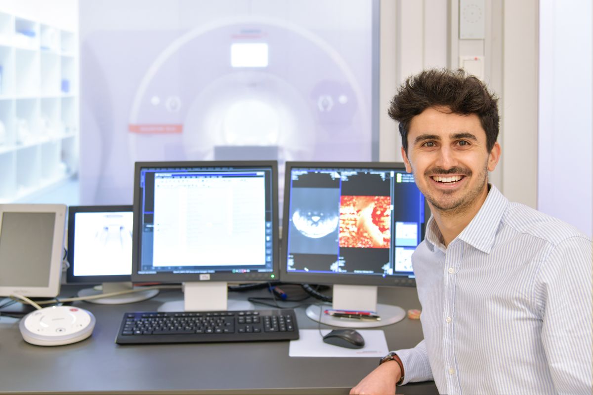
This is the area Dr. Simon Lévy specialises in, and for which he has recently received the prestigious scholarship from the Alexander von Humboldt Foundation. Thanks to the funding, he can now develop new procedures for obtaining MRI images of the spinal cord at the Institute of Radiology at Universitätsklinikum Erlangen, where he is involved in the working groups Metabolic and Functional MRI Imaging and the Digital Health Data Center. He uses cutting-edge MRI systems with magnetic field strengths of up to 7 tesla that show up the smallest structures in the spinal cord.
The MRI works on the basis of a strong magnetic field and radio waves, with the strength of the magnetic field being given in tesla. Whilst MRI systems with 1.5 and 3 tesla are already commonly used in clinical practice, the ultra high field MRI with a strength of 7 tesla – equivalent to 140,000 times the strength of the Earth’s magnetic field – has also been approved for use in recent years. Scanners with this flux density make even the finest structures and metabolic processes in tissue visible.
‘My research focuses on how information from the 7 tesla ultra high field MRI can be used to improve the image quality of the more commonly used 1.5 or 3 tesla MRI systems or to reduce scanning times,’ explains Dr. Simon Lévy. ‘In addition, my team and I are also developing techniques for investigating circulation in the spinal cord without using contrast agents, as not all patients tolerate them well, and the products incur additional expenses for the health system.’
Excellent research environment
Simon Lévy believes that Erlangen offers unique conditions for his project, unlike anywhere else in the world: ‘Erlangen is an amazing location for MRI research. The local institutions, the excellent equipment, the ongoing interaction with doctors and collaboration with Siemens Healthineers AG, where the systems are developed: better structures are nowhere to be found. I also really appreciate our team spirit. I arrived in Erlangen at the time of the coronavirus pandemic. In spite of being in a lockdown, my colleagues helped me get off to the best possible start,’ enthuses the 31 year old, whose career to date has included studying maths and physics in Paris and biomedical engineering in Montreal. He obtained his doctoral degree at Aix-Marseille University with research into using 7 tesla MRI scans to investigate the spinal cord.
Another advantage in Erlangen is that Universitätsklinikum Erlangen can offer all clinically approved whole-body MRI field strengths of 0.55, 1.5, 3 and 7 tesla. ‘That is perfect for our comparative studies. A stronger field strength provides a higher signal-noise ratio, but requires more advanced clinical infrastructure and entails more technical challenges and additional exclusion criteria for patients,’ explains Dr. Lévy.
In particular, the newly installed high performance MRI scanner with a low field strength of 0.55 tesla offers several practical benefits. The fact that it incurs comparatively low production and maintenance costs and does not need any powerful infrastructure also makes it particularly suitable for developing regions or remote areas. ‘We hope to use super high resolution deep learning methods to allow us to improve the quality of scans taken at 0.55 tesla on the basis of the improved image quality provided by 7 tesla. This solution would allow us to achieve a high-quality diagnostic image with a reasonably priced MRI system.’
About the Alexander von Humboldt Foundation
Today’s Alexander von Humboldt Foundation was founded in 1953 with the aim of promoting international collaboration in research, encouraging scientific progress and finally strengthening Germany’s position as an exceptional location for science and research. The Alexander von Humboldt Foundation awards the Humboldt research fellowship to support outstanding researchers from across the globe.
Further information
Dr. Simon Lévy
Phone: +49 9131 8525557