New microscope for materials research
FAU awarded 3.1 million euros in funding for high-resolution X-ray microscope
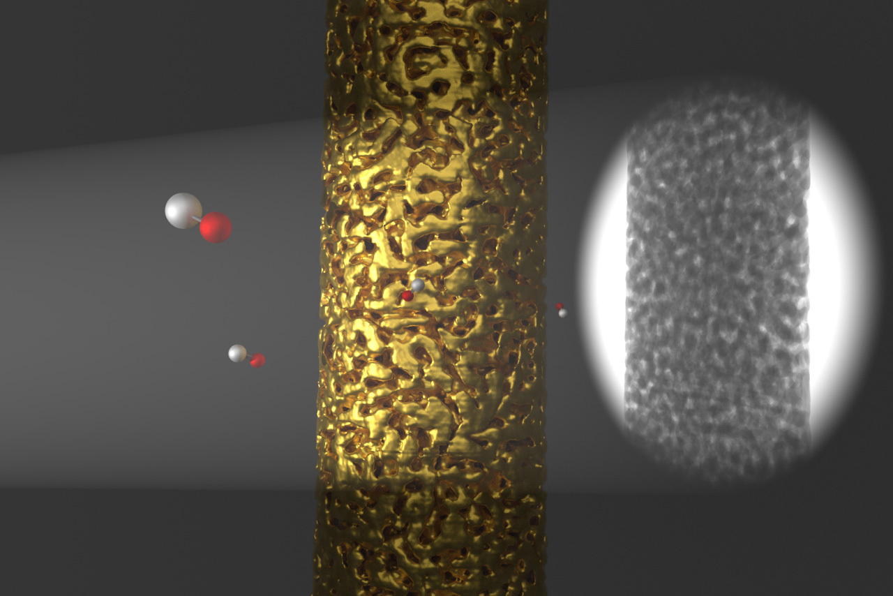
The Center for Nanoanalysis and Electron Microscopy (CENEM) at FAU has been granted funding for a high-resolution X-ray microscope that will allow its materials scientists to take their research in a completely new direction. In the future the researchers will use the microscope to conduct non-destructive analyses of the 3D structure of complex materials on a sub-micrometre scale and use the information obtained for the targeted development of new materials. This technology makes it possible to explore the tiny pore structure of nanofoams and optimise them for catalytic applications, for example. The X-ray microscope can also make minute cavities and cracks inside materials visible, enabling the early stages of material failure to be studied.
The German Research Foundation (DFG) is investing a total of 13.4 million euros in the establishment of this new technology at outstanding research centres – around 3.1 million euros of which have been allocated to FAU. The University was awarded the funding after a highly competitive application procedure involving five other institutions. ‘The funding is confirmation of the strength of materials research in Erlangen and of the University’s targeted investment in the field of modern materials analysis,’ says Prof. Dr. Erdmann Spiecker, Chair of Micro and Nanostructure Research, who submitted the application for CENEM. ‘In combination with CENEM’s other excellent microscopes, the new microscope will allow us to carry out multiscale materials characterisation, making us the only research centre in Germany to conduct this kind of research.’ Research on hierarchical structures, a focus of materials research at FAU and of the Cluster of Excellence ‘Engineering of Advanced Materials’, will benefit particularly significantly from the new technology. The combination of multiscale characterisation and modelling is an especially important part of this work.
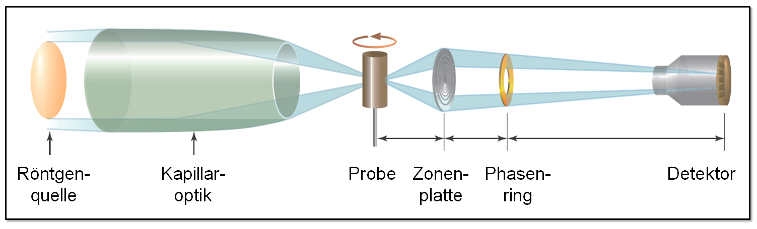
The Erlangen-based DFG Research Training Group GRK 1896 ‘In situ microscopy with electrons, X-rays and scanning probes’, in which over 20 doctoral candidates are researching modern materials testing and characterisation methods for small scales, will also benefit considerably from the new X-ray microscope. The microscope is equipped with a micromechanical testing unit that enables microscopic samples to be deliberately deformed and tomographic analyses to be carried out. ‘This will allow us to gain information on deformation mechanisms inside samples that we were not able to access before,’ Prof. Spiecker explains.
The new microscope will also be used as a basis to intensify long-term collaboration with the Development Center for X-ray Technology (EZRT), part of the Fraunhofer Institute for Integrated Circuits (IIS). EZRT develops efficient methods and systems using X-ray and optical technologies in the field of non-destructive testing that enable materials to be characterised and components to be tested without damaging them. The planned collaborative research between ERZT, CENEM and FAU’s materials scientists will make use of a wide range of synergies that result from the participants’ different expert knowledge and the different experimental possibilities, as well as the combination of basic and applied research.
Weitere Informationen:
Prof. Dr. Erdmann Spiecker
Phone: +49 9131 8528603
erdmann.spiecker@fau.de
cenem.fau.de