How solar cells could help find bone fractures
FAU researchers use new material for X-ray detectors
It is not about sunlight but X-rays: FAU researchers, together with colleagues from Austria and Switzerland, have demonstrated for the first time that a type of semiconductor that is normally used in solar cells is also sensitive to X-rays and reacts just as well as the materials that have been used for this purpose until now. The advantages of this are that these semiconductors are much simpler to produce, making them considerably less expensive, and that they can be used for different applications. The researchers recently published their findings in the journal Nature Photonics*.
While X-rays were recorded on photographic film in the past – and sometimes still are today – we now have digital X-ray cameras. These cameras, however, are very expensive. The detectors have semiconductors made of silicon or selenium which are installed in them using complex and time-consuming vacuum techniques. Meanwhile, a type of semiconductor known as a solution-processed semiconductor is being used more and more frequently in solar technology which, like X-ray technology, is essentially about absorbing light. These semiconductors can easily be printed onto substrates such as glass or plastic, helping to save a great deal when it comes to production costs. A team of FAU researchers led by Prof. Dr. Wolfgang Heiß at the Chair of Materials for Electronics and Energy Technology, together with researchers at Johannes Kepler University Linz and ETH Zürich, investigated whether these semiconductors which react to light waves in solar cells could also be suitable for X-ray imaging.
To take an X-ray image of bones in the body, X-rays are directed at the affected area. The X-rays penetrate the body where they are absorbed to a greater or lesser extent by different kinds of tissue and then captured by the detector behind it. The detector must absorb a high amount of the X-rays that reach it in order to obtain a usable image.
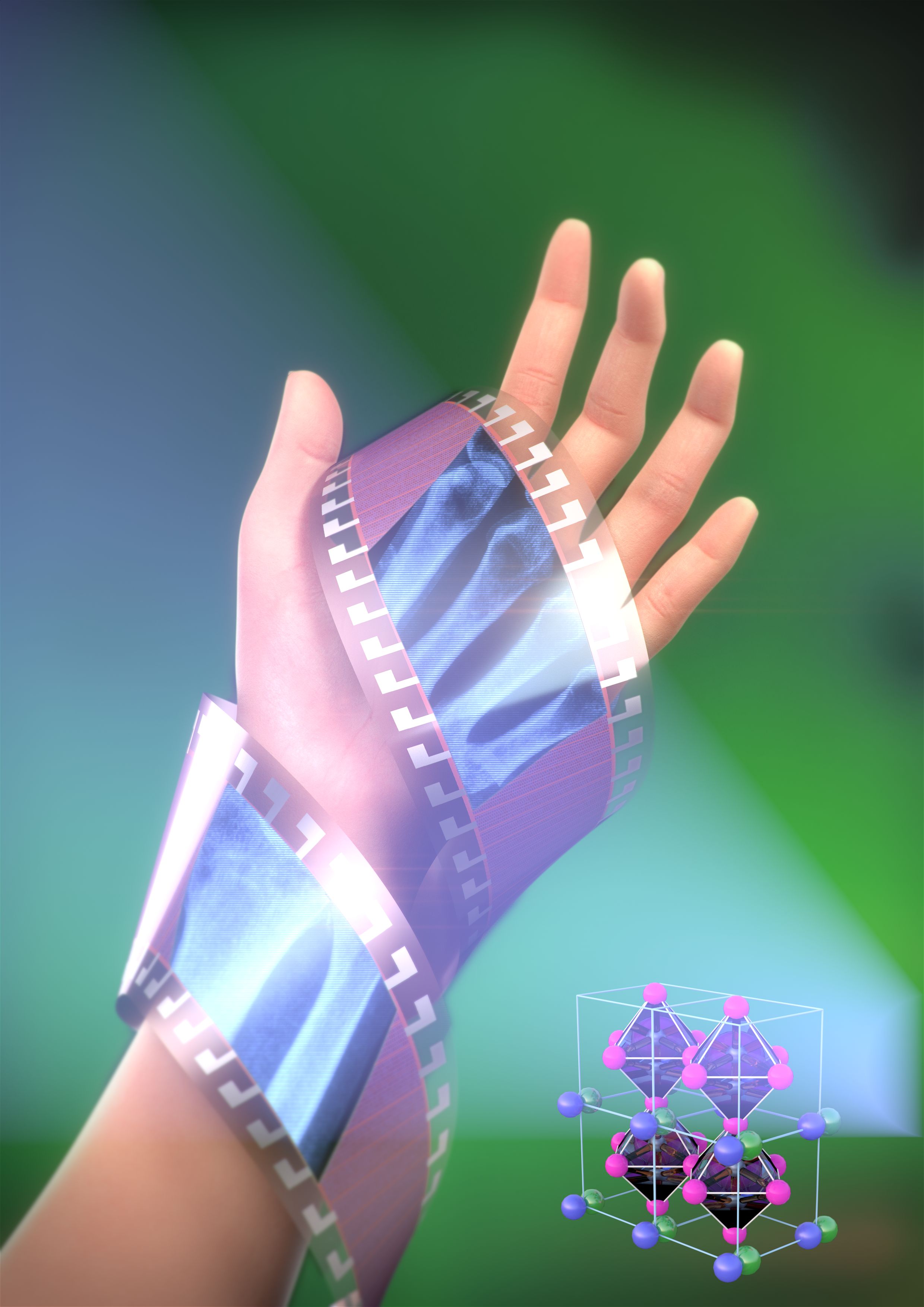
In their experiments the researchers directed X-rays at a thin-film solar cell consisting of a semiconductor made from metal-organic perovskites. This semiconductor contains a high amount of lead, among other materials. Lead – unlike lighter elements or biological substances – absorbs X-rays relatively well. The researchers determined that in principle this type of semiconductor is well suited to detect X-rays. However, they discovered that the solar cells themselves are far too thin to be used as detectors. They are only a few millimetres thick, meaning that they do not absorb enough X-rays and therefore the images that they create are not sharp or detailed enough. In response to this their colleagues at Johannes Kepler University Linz created detectors in which the semiconductor layer was around 300 times as thick as in the solar cells. The researchers applied the materials using airbrush technology, allowed them to create a homogeneous layer with sufficient electrical properties to detect X-rays.
They were particularly surprised to find that the results that they obtained with the semiconductors based on the solar cell technology were just as good as those produced by detectors which use solid-state semiconductors, which require complex vacuum deposition systems to produce them. The researchers’ results are therefore a big step towards the development of less expensive X-ray detectors. In the future the semiconductors could be sprayed onto flexible plastics such as Kapton or PET which would mean that they could be used in many more different areas for material and medical analysis. Airbrush technology could be particularly suitable for large-area detector arrays that are used, for example, in X-ray cameras used in medical technology for coronary angioplasties, or in materials testing.
*doi:10.1038/nphoton.2015.82
Further information:
Prof. Dr. Wolfgang Heiß
Phone: +49 911 568549216
wolfgang.heiss@fau.de