Endomicroscopy: diagnosing tissue without artificial contrast agents
A number of diseases can only be diagnosed by doctors taking and analysing tissue samples under the microscope. FAU researchers have now developed a procedure which allows tissues to be analysed in accessible body cavities without surgical intervention or the need to apply dyes intravenously or locally, for example on the intestinal mucous membrane. ‘Integrating multi-photon microscopes into endoscopes could allow doctors to assess the degree of inflammation in real time,’ says Prof. Oliver Friedrich from the Chair of Medical Biotechnology. ‘At the same time, it simplifies the procedure as no external dyes are required.’
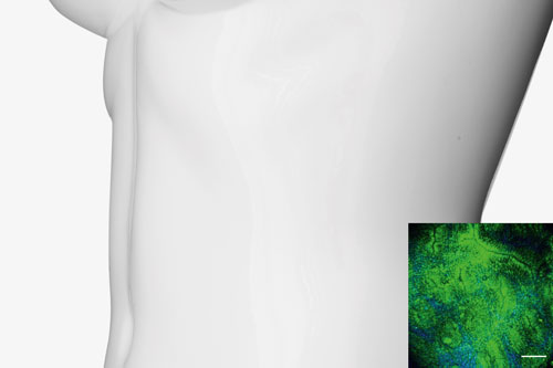
The researchers hope that the new procedure which provides detailed 3D images at a resolution on the micrometre scale will allow doctors to see below the surface of the intestine and detect even the slightest changes to cells in the organ or parts of the cell wall. ‘In future, the procedure could supplement biopsies or perhaps even make them superfluous in some cases.’
Multi-photon microscopy is not new. It has been used in fundamental research since the 1990s. A multi-photon microscope emits focused laser pulses at very high intensity for an extremely short period of time which are focused by the lens in the microscope. During this process, two or more photons hit certain molecules in the body simultaneously which then makes the molecules illuminate. The wavelength (colour) of the light which is transmitted allows conclusions to be drawn concerning the composition of the tissue.
As the technique provides three-dimensional images with an optical resolution of just a few micrometres, operators can see inside the tissue to a depth of several hundred micrometres. Multi-photon microscopes are already in use in medical applications, especially for examining the surface of the skin. Dermatologists, for example, use them to look for malignant melanoma.
By integrating this technique into an endoscope, doctors will in future be able to see inside the living human body, for example the intestinal wall. The Chair of Medical Biotechnology has been systematically analysing tissue samples for many years to track how tissue structures change as diseases progress or during the ageing process. In combination with information provided in real time from a multi-photon microscope integrated into an endoscope, this knowledge could provide new options for early diagnosis and treatment of many different diseases. ‘That is the vision driving our research,’ says Friedrich.
The FAU researchers have now taken a major step forward. They have succeeded in integrating the entire microscope technology including mini rod lenses into an endoscope. The objective lens is housed in a pin-sized cannula that is 32 millimetres long and has a diameter of 1.4 millimetres. The focal point can be adjusted electronically to vary optical penetration. The prism located on the point of the endoscope allows a sideways view into the colon and means that various rotational images of the tissue can be made from the same position.
The technique is already being tested in preclinical trials. At the moment, however, a rigid system is being used to transmit the visual information. The anatomy of the colon with its bends and curves means that rigid systems are not suitable for use in human intestines. ‘In order to use a flexible endoscope, we will have to integrate the entire scanning mechanism directly into the endoscope, not just the lens,’ explains Dr. Sebastian Schürmann, who is leading research in this area. ‘We still have some research to do.’
by Frank Grünberg
Here we present four further imaging methods:
Endomicroscopy: Rapid test for polyps
Computer tomography: weight-bearing scans
Light-sheet microscopy: an underground train system for immune cells
Multi-spectral optoacoustic tomography (MSOT): tracking haemoglobin
FAU research magazine friedrich
 This article first appeared in our research magazine friedrich. You can order the print issue (only available in German) free of charge at presse@fau.de.