FAU’s Uniklinikum Erlangen involved in collaborative project on the routine use of deep learning algorithms
Algorithms from artificial intelligence (AI) are being used more and more frequently, also for medical diagnosis. However, their potential is barely being tapped in a number of areas. A collaborative project from Universitätsklinikum Erlangen (UKER) at Friedrich-Alexander-Universität Erlangen-Nürnberg (FAU) and Gravina Hospital in Caltagirone (Italy) is showing that it does not need to be that way. The researchers are demonstrating how AI can be seamlessly integrated into clinical practice in a fully digitized department of pathology. Their findings have now been published in the journal Genome Medicine.*
Each year, more than 1.4 million people in Germany are treated in hospital for cancer. When a tumor is surgically removed, the tissue is usually examined in the department of pathology: which type of cancer is it exactly? Is the growth malignant? Should chemotherapy be offered, and if so, with which medication?
AI algorithms can help pathologists find the answers to these and other questions, for example by highlighting malignant transformation in digitalized tissue samples. However, their full potential often still remains untapped today. This is due in part to examination methods: while an MRI or ultrasound scan can produce digital images that can be assessed directly using AI, that is not the case with a tissue sample. “Until now, samples have mainly been examined using microscopes,” explains PD Dr. Fulvia Ferrazzi who leads the working group for bioinformatics and computer-assisted pathology at the Department of Nephropathology (head: Prof.Dr. Kerstin Amann) and at the Institute of Pathology (director: Prof. Dr. Arndt Hartmann) at UKER. “Digitalizing histopathological samples to obtain high-resolution images remains an exception.”
The Department of Pathology (director: Dr. Filippo Fraggetta) at Gravina Hospital in Caltagirone in Italy is already a step ahead – they routinely digitalize all tissue samples. “The problem here is not the availability of digital data,” comments Miriam Angeloni, who is pursuing a doctoral degree in Ferrazzi’s working group. “Rather, there has been no way of analyzing these data automatically using deep learning models until now.” This is the reason why AI tools are not yet routinely integrated into clinical diagnosis. “We investigated how we could integrate the use of these tools more smoothly.”
How does a fully digitalized department of pathology work?
When a tissue sample arrives in the pathology laboratory in Gravina Hospital, it goes through several processing steps. As a rule, several extremely thin specimens are prepared, fixed on thin glass slides and dyed with various chemicals. Next, high-resolution digital images are produced of these slides. Employees can access these images directly via the laboratory information system (LIS). The diagnosis is then made not like normal using a microscope but on a computer screen instead.
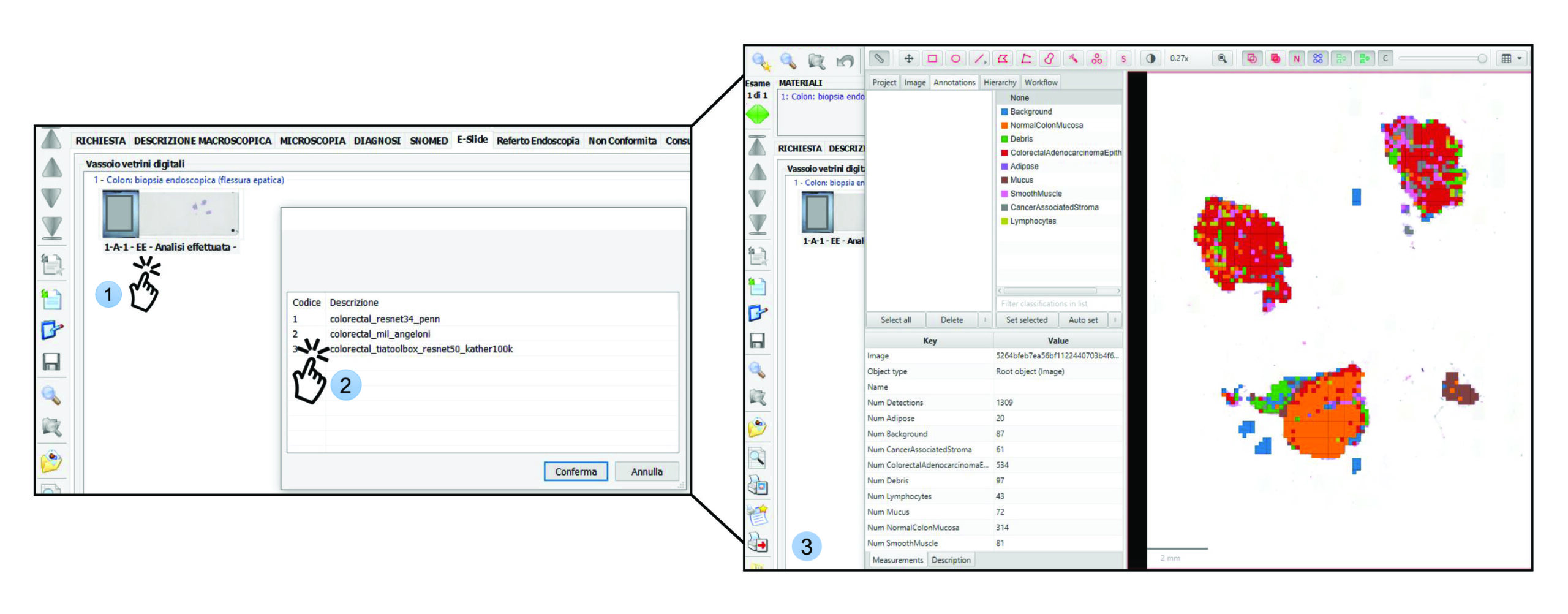
During their collaborative project, the researchers have developed a procedure that automatically integrates AI analysis into their workflow: As soon as new scans are entered in the LIS, all information required for the analysis is automatically transferred to a server with various AI models. There, the suitable algorithms are selected depending on the dyeing method that was used and the tissue from which the sample was taken. In addition to this standard procedure, the pathologists are also able to select an “on demand” analysis directly from the LIS.
Improved integration is hoped to improve the accuracy of the algorithms
The results of the analysis are then returned to the LIS. There, the algorithms’ predictions can be shown as “heatmaps”. These colored superimpositions can be used, for example, to indicate malignant regions on the digitalized tissue sample.
“Together with our collaboration partners we hope to use the workflow we have developed to provide clinical validation of the integrated deep learning models,” explains Fulvia Ferrazzi. The aim is to continue to improve the algorithms’ accuracy in future. “We also hope that our collaboration project will encourage the integration of deep learning models into routine diagnostics for other departments of pathology.”
*DOI: https://doi.org/10.1186/s13073-025-01484-y
To the original publicationFurther information:
PD Dr. Fulvia Ferrazzi
Working group for bioinformatics and computer-assisted pathology
Phone:09131/85-43677
Email: fulvia.ferrazzi@uk-erlangen.de
Miriam Angeloni
Working group for bioinformatics and computer-assisted pathology
Phone: 09131/85-48183
Email: miriam.angeloni@uk-erlangen.de