
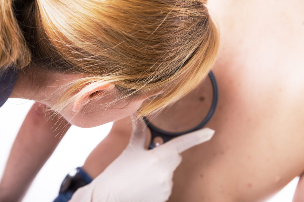
Researchers at FAU combine optical diagnosis method with AI-assisted image recognition.
Until now, diagnosing skin tumors has relied on detecting visible changes to the skin. Line-Field Confocal Optical Coherence Tomography (LC-OCT) is a well-established method used for diagnosis. Now, researchers at Friedrich-Alexander-Universität Erlangen-Nürnberg (FAU) led by Dr. Moritz Ronicke from the Department of Dermatology at Uniklinikum Erlangen have developed a new approach: In a feasibility study, the team introduced a procedure involving systematic screening of the faces of patients at a high risk of developing skin cancer. Using this procedure, researchers succeeded for the first time in detecting basal cell carcinoma even before any visible changes to the skin developed.
“Detecting basal cell carcinoma early allows for less invasive treatment options.”Dr. Moritz Ronicke
Basal cell carcinoma
Basal cell carcinoma is the most common type of skin cancer across the globe. It spreads locally, destroying the skin in its vicinity and thereby potentially damaging structures such as the nose or eyes if it occurs on the face. “Detecting basal cell carcinoma early allows for less invasive treatment options,” claims Dr. Moritz Ronicke. “In some cases, all that is required is a cream.”
LC-OCT delivers high resolution, three-dimensional images of the skin.
LC-OCT combines two techniques: optical coherence tomography (OCT), that indicates the thickness and extent of the tumor, and confocal microscopy ((LC: Line-field Confocal Microscopy), that takes clear images at the cellular level. The result: vertical, horizontal and three-dimensional images of the skin in real time with a resolution at micrometer scale, in other words a thousandth of a millimeter. By way of comparison, a human hair is approximately 50 micrometers thick.
The role of artificial intelligence
The AI delivers in real time a color-coded probability value for the presence of basal cell carcinoma. This allows the physician to recognize suspicious areas at a glance. It is still the physician who makes the final diagnosis.
Future applications
LC-OCT is already being used as standard to detect basal cell carcinoma in the early stages and monitor the success of non-invasive therapies. “Our new approach using “SUBSCANS” would allow patients at a high risk of developing skin cancer to receive a diagnosis even earlier on,” explains Moritz Ronicke. “If the SUBSCAN is routinely used in future, this could lead to more widespread use of creams for treating basal cell carcinoma, even avoiding the need for surgery in such cases.” At the current time, however, it is not possible to offer SUBSCAN routinely, as there is still a lack of data on the sensitivity of the method and it is still too time-consuming.
Dr. med. Moritz Ronicke has been working at Uniklinikum Erlangen since 2020, and has been involved since May 2023 as a clinician scientist in the NOTICE program (“Networks of Tissue Responses in Inflammation and Cancer”) at FAU. The Department of Dermatology is working on integrating LC-OCT into clinical procedures and conducting research into how AI-assisted screening can increase the accuracy of diagnosis.
doi: 10.1001/jamadermatol.2026.2992
Further information:
Contact
Dr. med. Moritz Ronicke
Department of Dermatology at Uniklinikum Erlangen
moritz.ronicke@uk-erlangen.de